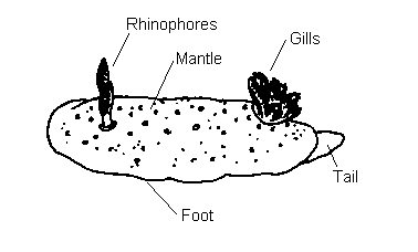
Anatomy
The mantle. In nudibranch molluscs the shell is only present in the larval stage. The loss of the shell in adults is probably responsible for the diversity of body forms present within this order.
|
In typical dorid nudibranchs, the mantle is thick and extends over the foot. The surface
of the mantle may bear tubercles which vary in size, shape and number and are often a character
used to identify nudibranchs. In many dorids acid glands and/or spicules are incorporated in the mantle tissue and it is thought that these are mainly defensive in function. However in Goniodorids, Polycerids and some other dorid families the mantle is progressively reduced to a ridge around the side of the body, from which pallial tentacles or processes arise. These processes usually have coloured tips and contain defensive glands and have been shown to produce chemicals distateful to fish. These chemicals are often manufactured from similar chemical compounds in the bryozoan or ascidian prey, or may be the same molecules selectively re-secreted by the nudibranch.
|
|
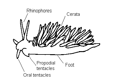
In aeolid nudibranchs the mantle is extended into long finger-like projections called
cerata (singular; ceras). The cerata contain branches of digestive gland and often this is
visible through the ceratal epidermis. In aeolids the tips of the cerata contain cnidosacs
which usually store nematocysts (stinging cells) that are obtained from ingested cnidarian
prey, such as hydroids, sea-anemones and soft corals. If disturbed, the nudibranch is capable
of discharging these stinging cells through a terminal pore in the ceras; this is an effective
deterrent to predatory fish.
|
The gills. In nudibranchs the gills are probably the most important respiratory organ, however gaseous exchange also occurs over the entire body surface. In dorids the gills consist of several feather-like structures that encircle the anus. This structure is termed the branchial plume and is situated in the posterior part of the animals' back. In true dorids (ie. Dorididae, Rostangidae, Chromodorididae, etc. ) the gills can be retracted into a gill-pocket. These dorids are known as cryptobranch dorids as opposed to phanerobranch dorids in which the gills are contractile but not retractile into a pocket. Goniodorids, Onchidorids and Polycerids are phanerobranch dorids. In aeolids the cerata function as gills. The ceratal epidermis is thin enough to enable oxygen from the surrounding water to diffuse in and carbon dioxide (a waste product of respiration) to diffuse out. In the dendronotaceans the cerata are branched or tuberculate, this increases the surface area available for gas exchange. The arminaceans are a mixed bag, with cerata in most species, occasionally branched, but in the Arminidae leaf-like gills are hidden in a ridge between the mantle and foot.
The oral veil. In some nudibranchs the front region of the head is extended to form an oral veil. This structure varies from one species to another e.g. in Dendronotus frondosus it has branched processes; in Polycera quadrilineata it forms up to six finger-like processes and in Doto species it is smooth edged with two lateral flaps.
The oral tentacles. Many nudibranch species have a pair of processes, one on either side of the mouth, which are probably involved in identifying food by taste or touch.